Tumoral Melanosis

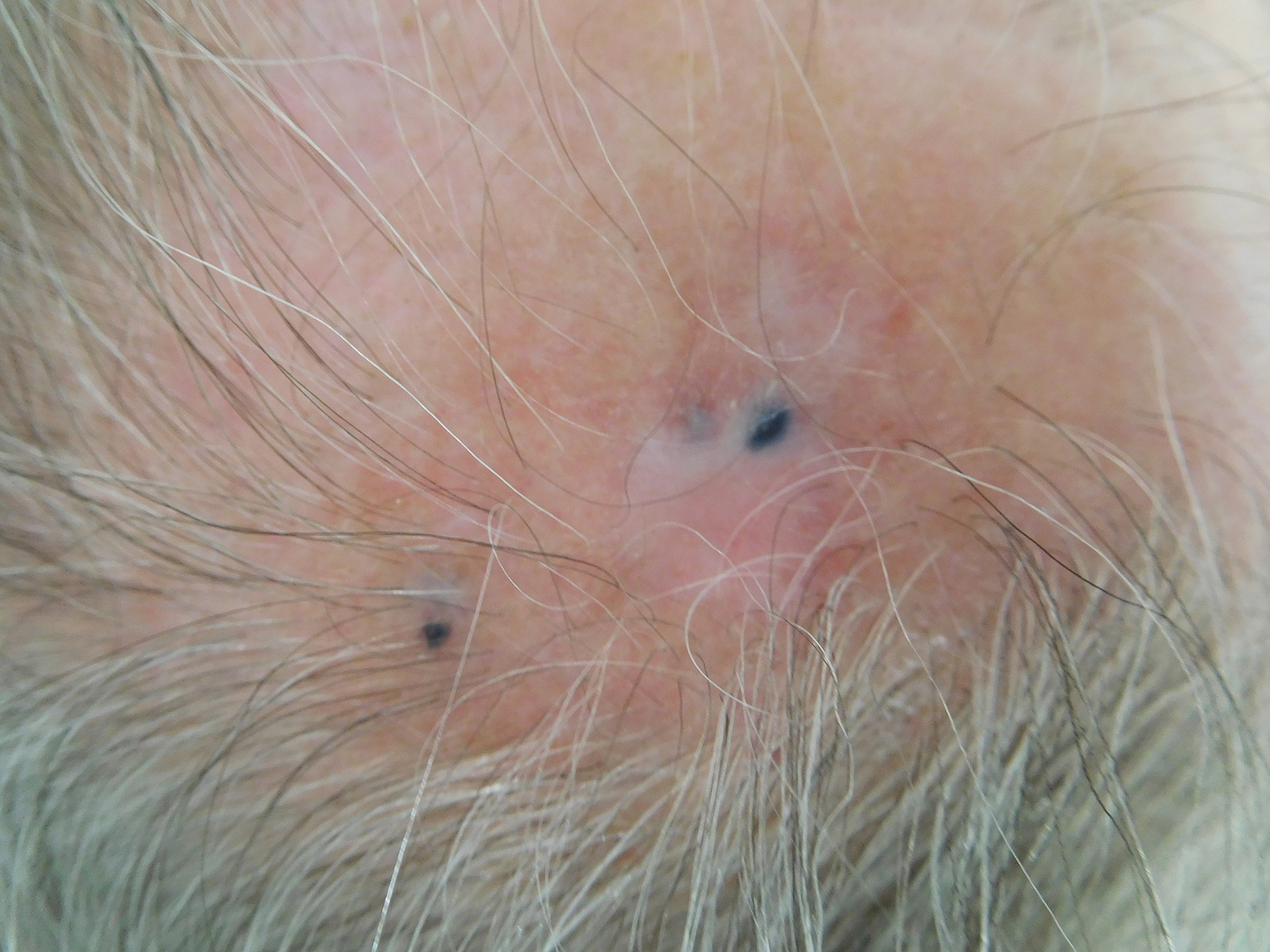
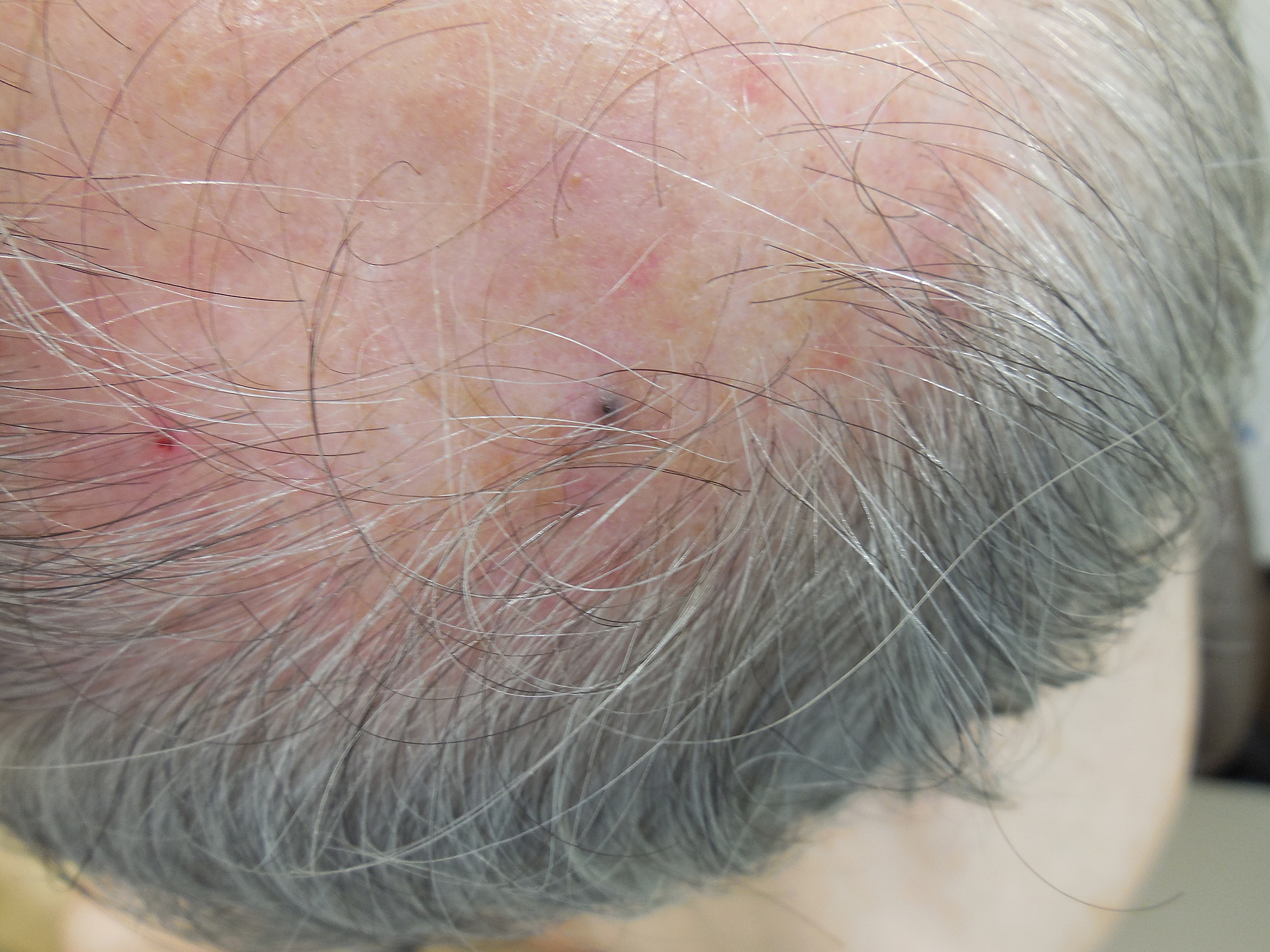
Tan et al reported a 69 year old indian man who presented with new onset vitiligo and cutaneous tumoral melanosis distant from the site of his primary melanoma 2.
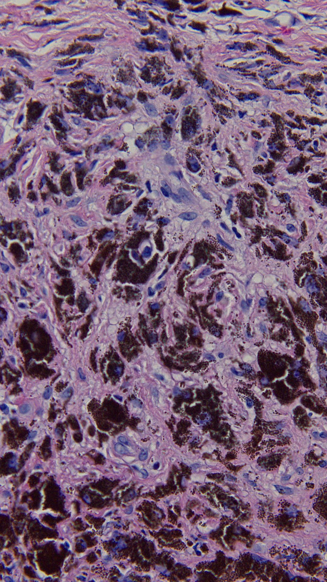
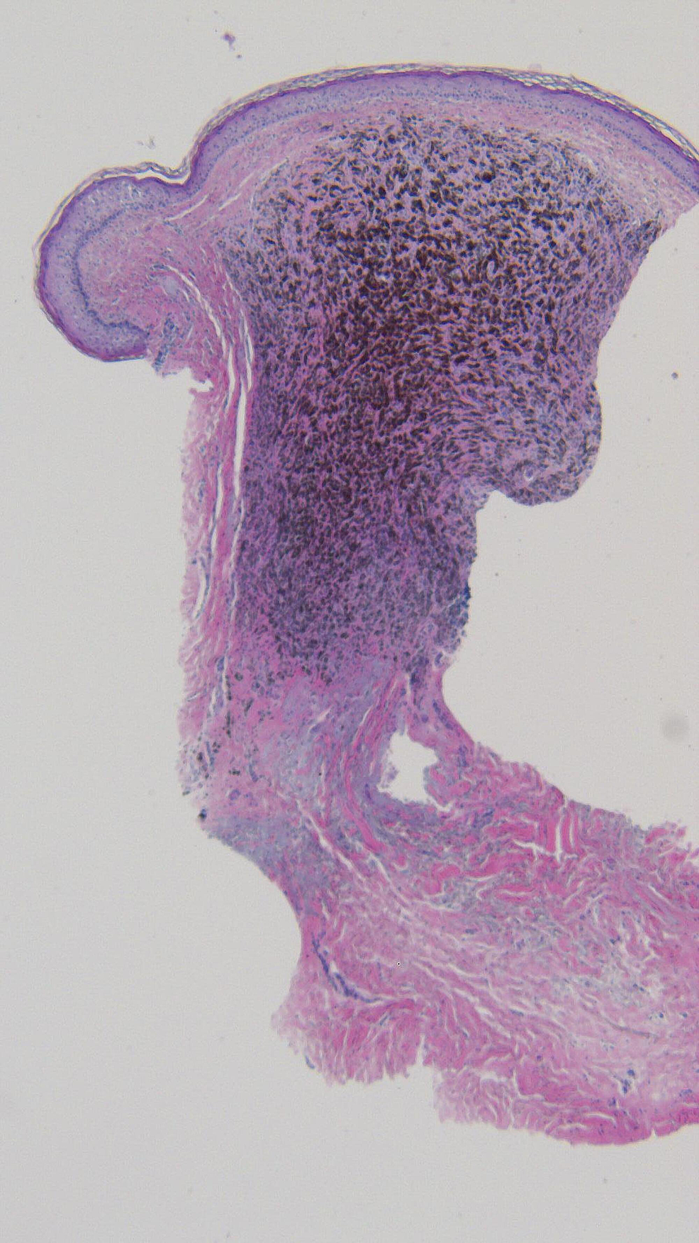
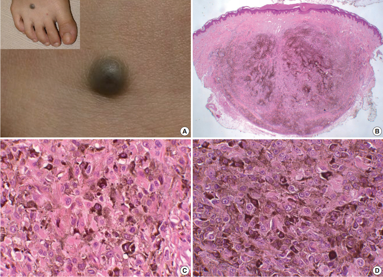
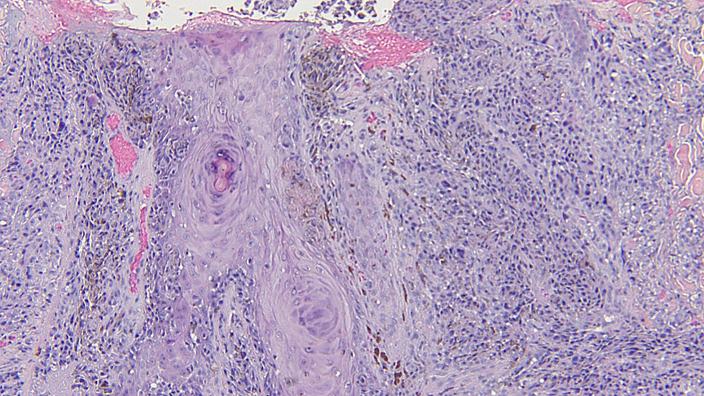
Tumoral melanosis. Experts think the lining of the colon darkens. Tumoral melanosis associated with pembrolizumab treated metastatic melanoma abstract. Melanosis coli is a harmless condition in which the lining of the colon and rectum which is usually pink in color turns a shade of black or brown. Tumoral melanosis is a histological term used to refer to a nodular accumulation of melanophages in the dermis which clinically arises as a pigmented lesion.
2019 new code 2020 billable specific code. Histology reveals dense dermal and subcutaneous. Abnormal dark brown or brown black pigmentation of various tissues or organs as the result of melanin or in some situations other substances that resemble melanin to varying degrees. 1 although partial regression is a relatively common finding in melanoma tumoral melanosis with complete absence of melanocytes is exceedingly rare.
For example melanosis of the skin may occur in widespread metastatic melanoma sunburn during pregnancy and as a result of chronic infections. Tumoral melanosis is a form of completely regressed melanoma that usually presents as darkly pigmented lesions. Tumoral melanosis clinically resembles metastatic melanoma occurs in the context of regressed disease and requires evaluation to rule out underlying melanoma and metastatic disease. It is usually associated with the.
Tumoral melanosis is a form of completely regressed melanoma that usually presents as darkly pigmented lesions suspicious for malignant melanoma. Other specified disorders of eye and adnexa. Histopathology demonstrates a nodular infiltrate of melanophages in the dermis subcutaneous tissue deep soft tissue or lymph. Interestingly mav has been reported to occur in tumoral melanosis a sign of complete regression of a primary mm in patients who present with metastatic melanoma.
